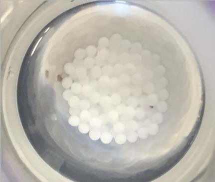
In a recent PNAS paper, researchers at MIT used a specific type of magnetic resonance imaging (MRI) to measure the oxygen levels of transplanted pancreatic islet cell particles in mice. Understanding how the levels of oxygen changed over time in mice can help engineers develop better methods of replacing particles of beta cells in humans. The study had funding from a few sources, including Breakthrough T1D, the Leona M. and Harry B. Helmsley Charitable Trust Foundation, the Parviz Tayebati Research Fund, and a Koch Institute Support Grant from the National Cancer Institute.
The researchers transplanted islet cells in particles made of alginate, a starchy natural material found in algae, into the intraperitoneal (IP) cavity of mice. This space between the lining of the abdominal cavity and the lining surrounding the body’s main organs is a promising place to transplant beta cells because from that location they can affect glucose levels in the body much more quickly. It is important that the implanted cells receive enough oxygen, which they need to stay alive and to keep producing insulin.
Previously, researchers had determined that bigger particles, those with a width of 1.5 millimeters, worked longer than smaller particles that were 0.5 millimeters wide. That had to do with the ability of the larger particles to avoid getting stuck together in areas that didn’t provide the necessary oxygen.
Once inside that IP cavity, it is difficult to track the cells, because they can move around freely. So, the scientists placed fluorine-containing materials into the particles. Then they used a fluorine MRI to measure oxygen levels in the IP space over a three-month period. Fluorine MRI can measure interactions between a magnetic field and fluorine nuclei, as well as how these interactions are affected by the presence of oxygen.
The scientists also measured the blood-glucose levels of the test and control mice in the study. To analyze the data more efficiently, the team used a machine-learning algorithm to go through all of the images and find links between the positions of the capsules within the IP cavity, oxygen levels, and the blood-glucose levels of the mice.
The analysis confirmed that smaller particles produce insulin for only about a month because by then they accumulate in fatty areas and fail to get the oxygen they need to keep going. Larger particles spread out over a larger area, and some of them hit upon high-oxygen areas. So they are able to secrete enough insulin to keep glucose levels stable over several months. The ultimate goal is to create little factories in the body that can supply insulin on demand for patients.
To learn more about how Breakthrough T1D is funding beta cell replacement therapies, visit here.